ECLIPSE Ti2
고급 이미징을 위한 최고의 플랫폼
ECLIPSE Ti2는 관찰 방법을 혁신적으로 변화시키는 독보적인 25mm 시야각(FOV)을 제공합니다.
이 놀라운 FOV를 통해 Ti2는 성능 저하 없이 대형 CMOS 카메라의 센서 영역을 최대화하며 데이터 처리량을 크게 향상시킵니다.
Ti2의 플랫폼은 매우 안정적이고 드리프트가 없도록 설계되어 초고해상도 이미징 요구 사항을 충족하며, 고유한 하드웨어 트리거 기능으로 매우 까다로운 고속 이미징 애플리케이션을 향상시키며,
또한 Ti2만의 지능적인 기능이 내부 센서에서 데이터를 수집하여 이미징 워크플로우를 통해 사용자를 안내하여 사용자에 의한 오류의 가능성을 제거합니다.
이미지를 획득하는 동안 각 센서의 상태가 자동으로 기록되어 이미징 실험의 품질을 관리하고 데이터 재현성을 향상시킵니다.
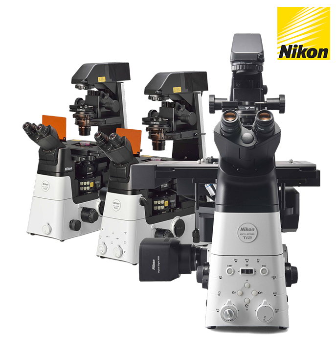
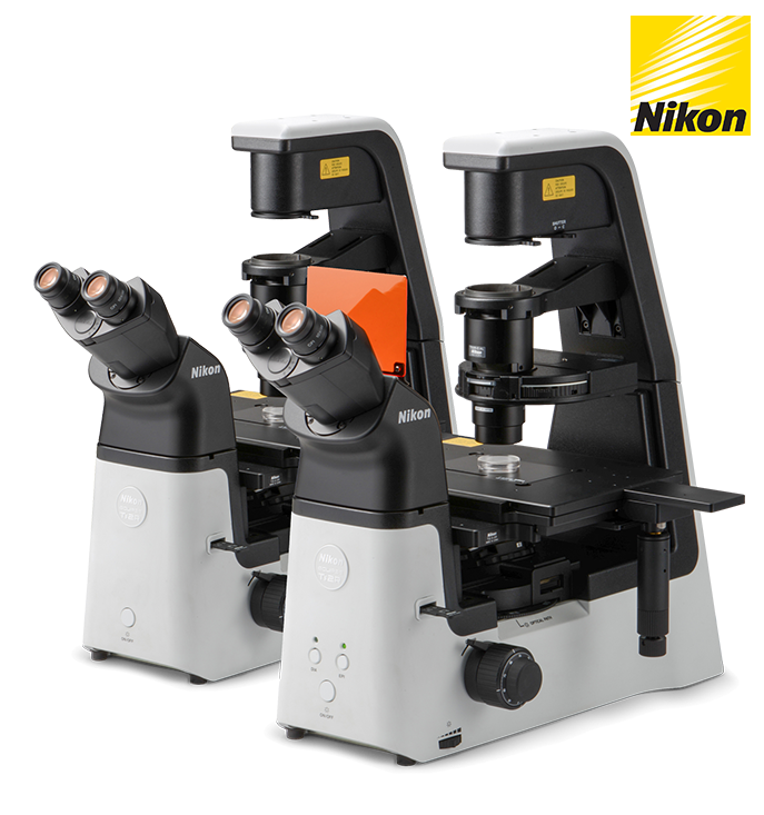
ECLIPSE Ti2 Series
-
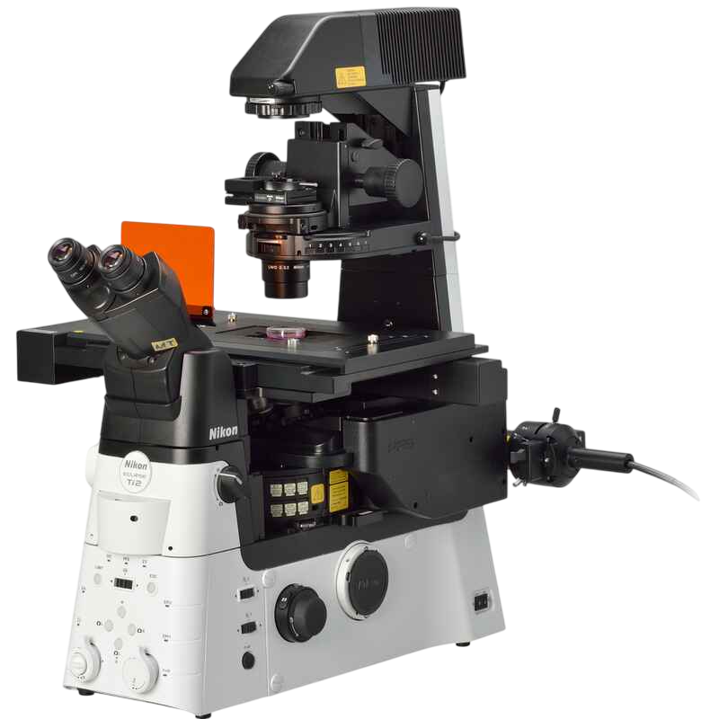
ECLIPSE Ti2-E
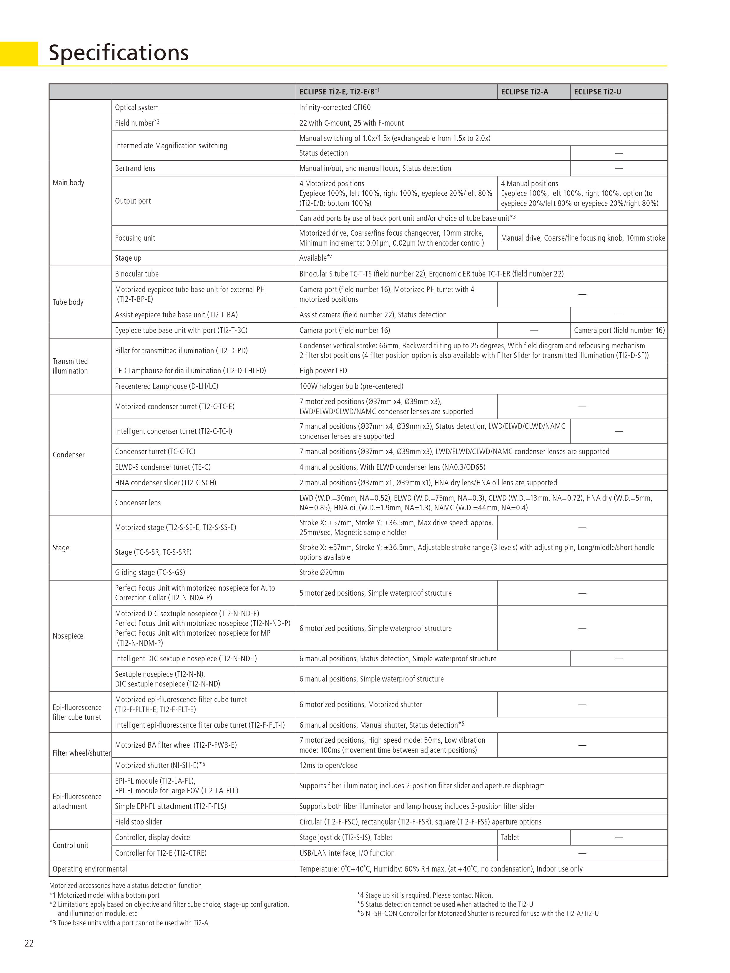
고급 이미징 애플리케이션을 위한 전동 지능형 모델. PFS, 자동 보정 칼라 및 외부 위상차 시스템과 호환됩니다. 살아 있는 세포 이미징, 고밀도 애플리케이션, 공초점 및 초해상도를 선택할 수 있는 기반이 됩니다. -
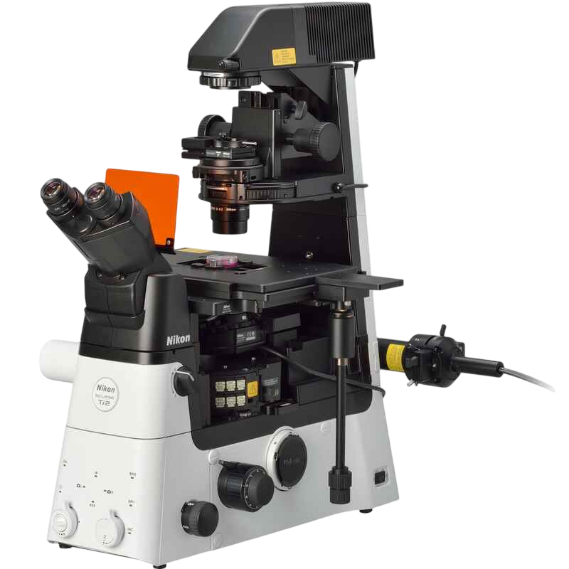
ECLIPSE Ti2-A
레이저 애플리케이션을 위한 이미징 기능을 갖춘 수동 모델. 지능형 기능은 이미징 워크플로우와 자동 현미경 상태 감지를 통해 대화형 지침을 제공합니다. -
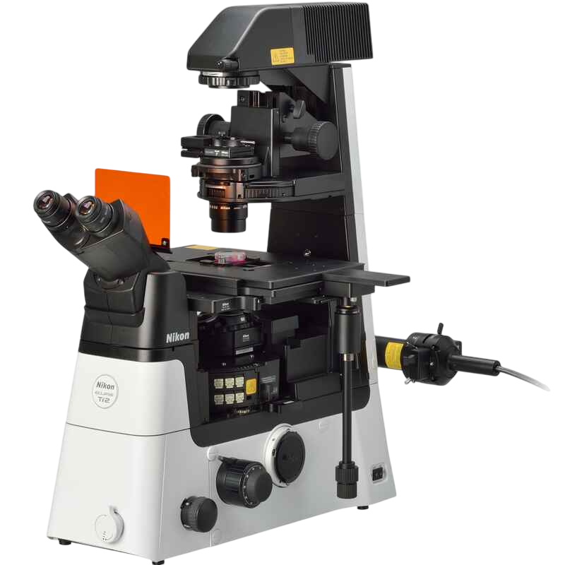
ECLIPSE Ti2-U
다양한 연구 용도에 적합한 기본 수동 모델입니다.
획기적인 FOV
-
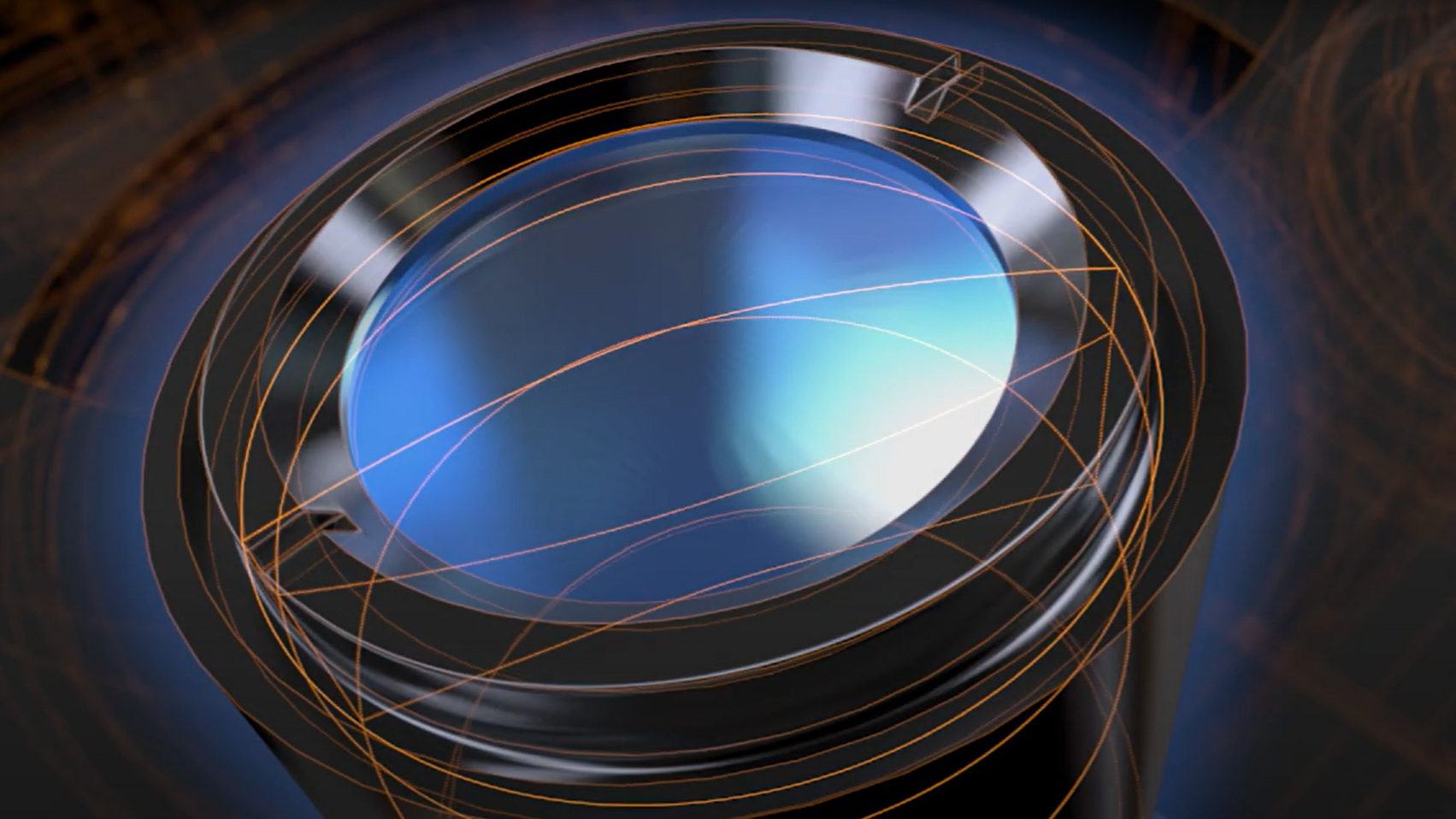
대규모, 시스템적 접근 방식으로 연구 추세가 발전함에 따라 더 빠른 데이터 수집과 더 높은 처리량에 대한 요구가 증가하고 있습니다.
대형 카메라 센서의 발달과 PC의 데이터 처리 능력 향상은 이러한 연구 경향을 촉진시켰습니다.
전례 없는 25mm 시야각을 갖춘 Ti2는 차세대의 확장성을 갖추었으며, 카메라 기술이 빠르게 발전하는 환경 속에서 연구자들에게 대형 검출기의 활용도를 극대화할 수 있는 미래 지향형 이미징 플랫폼을 제공합니다.
더 넓은 영역에 밝은 조명을
고출력 LED가 Ti2의 넓은 시야 전체에 밝은 조명을 제공하여 고배율 DIC와 같은 까다로운 애플리케이션에서 명확하고 일관된 결과를 보장합니다.
플라이아이 렌즈 디자인으로 스티칭 애플리케이션에서 고속 이미징의 정량적 분석과 이미지의 매끄러운 타일링을 위해 표면의 끝에서 끝까지 균일한 조명을 제공합니다.
-
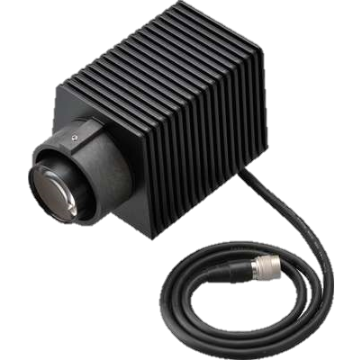
고출력 LED 조명
-
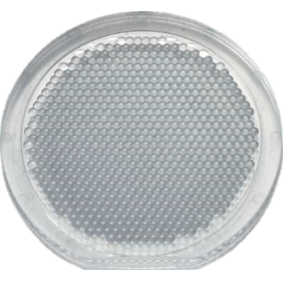
플라이아이 렌즈 내장
대형 FOV 이미징용으로 설계된 소형 에피 형광 조광기에는 쿼츠 플라이아이 렌즈가 있어 자외선을 포함한 넓은 스펙트럼에 걸쳐 투과율이 높습니다. 하드 코팅된 대구경 형광 필터가 신호 대 노이즈 비가 높은 큰 FOV 이미지를 제공합니다.
-
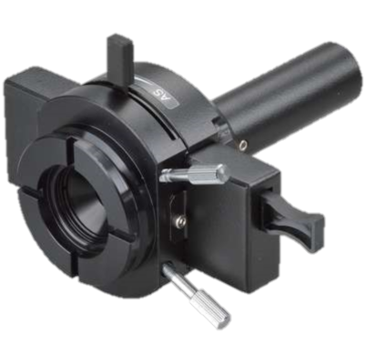
대형 FOV 에피 형광 조광기
-
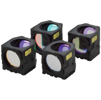
대구경 형광 필터 큐브
대용량 데이터 수집용 카메라
니콘의 FX 포맷 F-마운트 카메라 디지털 사이트 10 및 디지털 사이트 50M에는 전문 D-SLR 카메라용으로 개발된 연구 최적화 CMOS 이미지 센서가 장착되어 있습니다.
이를 통해 고속 및 고감도 라이브 셀 이미징이 가능하여 Ti2의 큰 FOV를 최대로 활용할 수 있습니다.
대구경 관찰 렌즈
이미징 포트에서 25가지 필드를 실현하고자 관측광로의 직경이 확대되었습니다.
결과적으로 커진 FOV가 기존 광학 영역의 약 두 배를 캡처할 수 있어 CMOS 검출기와 같은 대형 센서를 통해 최대의 성능을 얻을 수 있습니다.
-
튜브 렌즈 확대
-
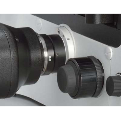
25가지 큰 필드 번호가 있는 이미징 포트
큰 FOV 이미징을 위한 대물렌즈
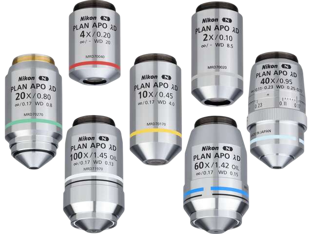
우수한 이미지 평탄도의 대물렌즈가 표면의 끝에서 끝까지 고품질 이미지를 보장합니다.
OFN25 대물렌즈의 능력을 최대로 활용하여 데이터 수집을 크게 가속화합니다.
대용량 데이터 수집용 카메라
니콘의 FX 포맷 F-마운트 카메라 디지털 사이트 10 및 디지털 사이트 50M에는 전문 D-SLR 카메라용으로 개발된 연구 최적화 CMOS 이미지 센서가 장착되어 있습니다. 이를 통해 고속 및 고감도 라이브 셀 이미징이 가능하여 Ti2의 큰 FOV를 최대로 활용할 수 있습니다.
타의 추종을 불허하는 니콘 광학 장치
다양한 기법의 정교한 관찰에 사용하도록 설계된 니콘의 고정밀 CFI60 인피티니 광학은 뛰어난 성능과 견고한 신뢰성으로 연구원들에게 높은 평가를 받고 있습니다
아포다이즈 위상차
선택적 진폭 필터가 적용된 니콘 고유의 아포다이즈 위상차 대물렌즈는 대비를 극적으로 높이고 후광 효과를 줄여 상세한 고화질 이미지를 제공합니다.
-
아포다이즈 위상판이 APC 대물렌즈에 통합되어 있습니다.
-

CFI S Plan 플루오 ELWD ADM 40XC 대물렌즈로 캡처한 BSC-1 세포
외부 위상차
Ti2-E
전동식 외부 위상차 시스템이 있어 사용자는 위상차 대물렌즈 없이도 형광 전송을 손상시키지 않고 위상차와 에피 형광 이미징을 결합할 수 있습니다.
예를 들어, NA가 매우 높은 담금 대물렌즈를 위상차 이미징에 사용할 수 있습니다.
이러한 외부 위상차 시스템을 사용하여 사용자는 TIRF 및 레이저 핀셋 애플리케이션과 같은 약한 형광 이미징을 포함한 다른 이미징 양식과 위상차를 쉽게 결합할 수 있습니다.
-
① 위상 링이 내장된 아이피스 베이스 유닛
② 위상 링
③ 위상 링이 없는 대물 렌즈 -

반사 형광 및 외부 위상차 이미지
CFI Apochromat TIRF 100XC Oil 대물렌즈 로 캡처한 GFP-alpha-tubulin으로 표지된 PTK-1 세포
사진 제공
DIC(미분 간섭 대비)
높은 평가를 받는 니콘의 DIC 광학은 배율 범위 전체에서 높은 해상도와 대비로 균일하고 선명하고 상세한 이미지를 제공합니다.
DIC 프리즘은 각 대물 렌즈에 맞게 개별적으로 조정되어 모든 샘플에 대해 최고 품질의 DIC 이미지를 제공합니다.
-
개별 대물렌즈에 맞는 DIC 프리즘이 노즈피스에 장착됨
-
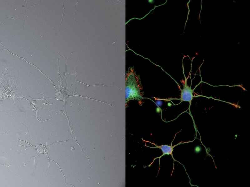
DIC 및 에피 형광 이미지:
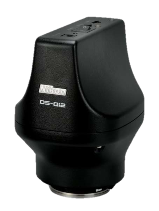
CFI Plan Apochromat Lambda 60XC 대물렌즈 및 DS-Qi2 카메라로 캡처한 뉴런(DAPI, Alexa Fluor® 488,
NAMC (Nikon Advanced Modulation Contrast)
난모세포와 같은 염색되지 않은 투명한 샘플을 위한 플 라스틱 호환 고대비 이미징 기술입니다. NAMC는 쉐도우 캐스트 모양의 유사 3차원 이 미지를 제공합니다. 대비 방 향은 각 샘플에 대해 쉽게 조 정할 수 있습니다.
-
NAMC 대물 렌즈에는 회전식 변조기가 포 함되어 있습니다.
-
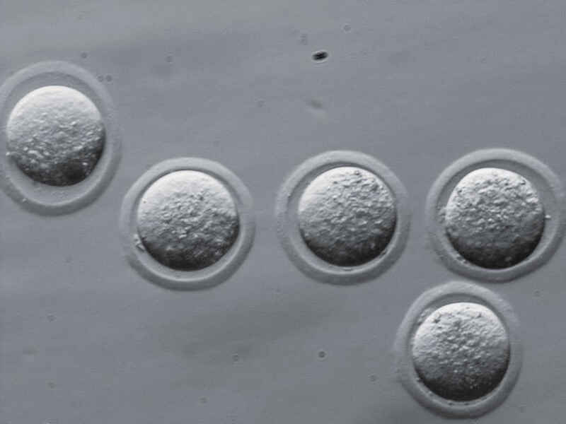
NAMC 이미지:
CFI S Plan Fluor ELWD NAMC 20XC 대물렌즈로 캡처한 마우스 배아
자동 보정환
Ti2-E
표본 두께, 커버 유리 두께, 표본의 굴절률 분포, 온도가 변하면 구면 수차와 이미지 열화가 발생할 수 있습니다. 최고 품질의 대물 렌즈에는 종종 이런 변화에 대해 보정하는 보정환이 있으며, 보정환의 정확한 위치는 고해상도, 고대비 이미지를 얻는 데 결정적으로 중요합니다. 이 새로운 자동 보정환은 하모닉 드라이브와 자동 보정 알고리즘을 이용하므로, 사용자는 보정환을 쉽게 정밀 조정하여 대물 렌즈의 성능을 매번 최적화할 수 있습니다.
-
보정환의 움직임을 매우 정확하게 제어하는 하모닉 드라이브 메커니즘
-

초고해상도 이미지(DNA PAINT): α-투불린(녹색) 및 TOMM-20(자홍색)을 발현하는 CV-1 세포를 CFI 아포크로마트 TIRF 100XC 오일 대물렌즈로 캡처한 이미지
에피 형광
니콘의 독점적인 나노 크리스털 코팅 기술을 활용하는 Lambda D 시리즈 대물렌즈는 넓은 파장 범위에서 높은 투과율과 수차 보정이 필요한 까다로운 저신호 다중 채널 형광 이미징에 적합합니다.
향상된 형광 감지 및 노이즈 터미네이터와 같은 미광 대책을 제공하는 새로운 형광 필터 큐브와 결합된 Lambda D 시리즈 대물렌즈는 단일 분자 이미징, 혹은 발광 기반 애플리케이션 등의 미약한 신호를 관찰할 때 그 힘을 보여줍니다.

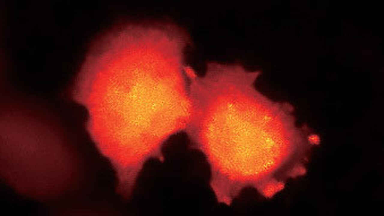
발광 이미지:
BRET 기반 칼슘 지표 단백질인 나노랜턴(Ca2+)을 발현하는 HeLa 세포.
샘플 제공: 나가이 다케하루 교수, 오사카 대학 과학 산업 연구소
새로운 부피 대비
Ti2-E
부피 대비 기법은 위상 분포 이미지를 조합하기 위해 다양한 Z-깊이에서 캡처된 일련의 무라벨 명시야 이미지를 이용합니다.
부피 대비는 세포를 자동 계산 및 면적 분석을 위한 물체로 쉽게 식별할 수 있도록 합니다. 이 방법은 명시야 이미징을 이용하므로, 세포의 인라인 비파괴 분석이 가능하여 폭넓고 다양한 응용 분야에 적합합니다..
참고. Ti2-E에만 해당.
CFI S Plan Fluor ELWD 20XC로 이미징한 HeLa 세포
-
Brightfield
-

Volume Contrast
부피 대비(VC)의 특징
무라벨 배양에서 세포를 정확하게 식별하여 세포 수 집계 및 면적 측정 자동화
-
Cell identification based on phase contrast images
Cells encircled in red are incorrectly identif -

Cell identification using VC images
Cells encircled in red are correctly identified as three sepa
메니스커스 효과가 세포 식별에 미치는 영향 최소화
메니스커스 효과는 위상차 이미지의 웰 가장자리에서 부정적인 영향을 미칩니다. 볼륨 대비 방법을 사용하면 이 효과를 피해 웰 가장자리에 있는 세포를 분명하게 식별할 수 있으므로, 집계되는 세포 수가 증가하고 통계가 개선됩니다.
-
Phase contrast image
-
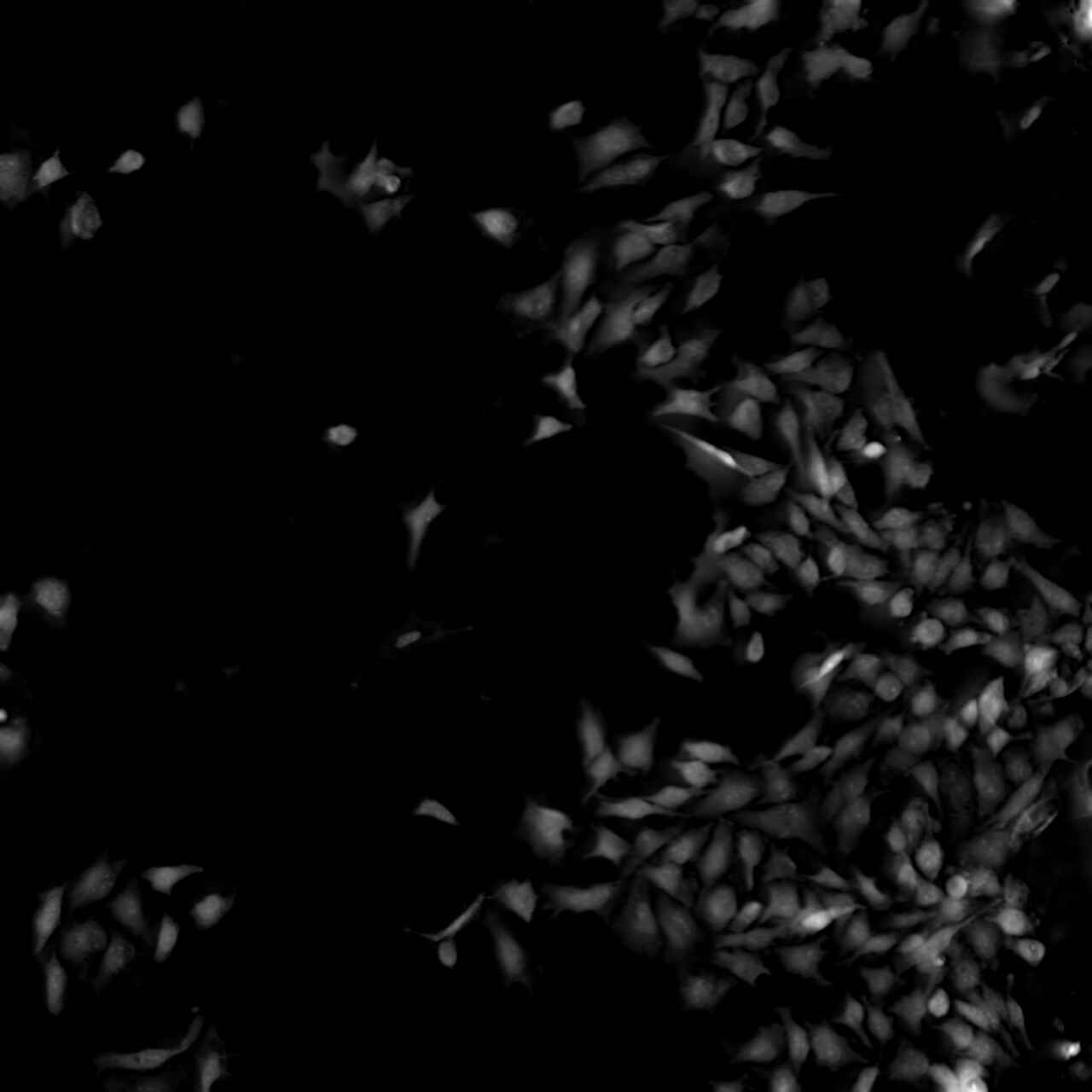
VC image
완벽한 초점
-

이미징 환경의 온도와 진동에 의한 아주 작은 변화도 초점 안정성에 큰 영향을 미칠 수 있습니다. Ti2는 정적 및 동적 측정을 모두 사용해 초점 드리프트를 제거하여 장시간 실험 중
매우 높은 안정성을 위해 기계적으로 재설계됨
Ti2-E
초점 안정성을 향상시키기 위해 Z 드라이브 및 PFS 자동 포커싱 메커니즘이 완전히 재설계되었습니다.
새로운 Z-포커싱 메커니즘은 진동을 최소화하기 위해 더 작고 노즈피스에 인접하게 배치됩니다. 확장(다단) 구성에서도 노즈피스에 인접하여 모든 적용에 대한 안정성을 보장합니다.
① 높은 안정성의 Z-포커싱 메커니즘은 확장된 구성에서도 노즈피스와의 인접성이 유지됩니다.
Perfect Focus System ( PFS ) 의 디텍터 부분은 대물렌즈 노즈피스의 기계적 부하를 줄이기 위해 노즈피스에서 분리되었습니다.
이 새로운 디자인은 또한 열 전달을 최소화하여 보다 안정적인 이미징 환경에 기여합니다. 이로 인해 Z-드라이브 모터의 전력 소비도 감소했습니다.
이러한 기계적 재설계과 결합하여 단일 분자 이미징 및 초고해상도 애플리케이션에 완벽하게 적합한 매우 안정적인 이미징 플랫폼이 탄생했습니다.
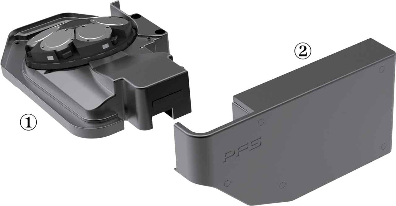
높은 안정성의 Z-포커싱 메커니즘은 확장된 구성에서도 노즈피스와의 인접성이 유지됩니다.
① PFS 노즈피스
② PFS 측정 단위
PFS를 통한 실시간 초점 보정
Ti2-E
PFS(Perfect Focus System)는 시료에 시약을 추가하거나 다중 포지션 이미징을 비롯한 다양한 요인으로 인해 발생할 수 있는 초점 드리프트를 자동으로 보정합니다.
PFS가 커버슬립 표면의 포지션을 실시간으로 감지하고 추적하여 초점을 유지합니다. 고유한 광학 오프셋 기술을 통해 사용자는 커버 슬립 표면에서 오프셋된 원하는 위치에 쉽게 초점을 유지할 수 있습니다. PFS는 내장된 선형 인코더와 고속 피드백 메커니즘을 통해 자동으로 지속적으로 초점을 유지하여 장기적이고 복잡한 이미징 작업 중에도 매우 안정적인 이미지를 제공합니다.
PFS는 플라스틱 배양 접시를 사용한 일상적인 실험부터 단분자 이미징 및 다광자 이미징에 이르는 광범위한 용도와 호환됩니다. 또한 자외선부터 적외선에 이르는 광범위한 파장과도 호환되므로 다광자 및 광학 족집게 용도에도 사용할 수 있습니다.
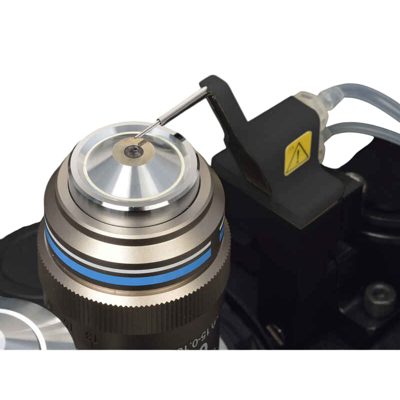
침지 분배기
Ti2-E
새로운 침지 분배기를 사용하면 담금 대물렌즈에 PFS가 결합되어 장기 이미징 성능을 높일 수 있습니다. 침지 분배기는 자동으로 적절한 양의 증류수를 대물렌즈 끝에 도포하여 실험 중에 침지액이 마르거나 넘치지 않도록 합니다. 모든 유형의 침수 대물렌즈와 호환되며 장기간에 걸쳐 고해상도, 고대비 및 수차 보정된 타임랩스 이미지를 안정적으로 제공하는 데 도움이 됩니다.
호환 대물 렌즈
CFI Apochromat LWD Lambda S 20XC WI
CFI Apochromat Lambda S 40XC WI
CFI Apochromat LWD Lambda S 40XC WI
CFI Plan Apochromat VC 60XC WI
CFI Plan Apochromat IR 60XC WI
CFI SR Plan Apochromat IR 60XC WI
CFI SR Plan Apochromat IR 60XAC WI
Assist Guide
더 이상 복잡한 현미경 정렬 및 작동 절차를 암기할 필요가 없습니다. Ti2는 센서의 데이터를 통합해 작동 단계를 안내하므로 사용자에 의한 오류를 방지하고 연구원이 데이터에만 집중할 수 있도록 합니다.

내장 센서는 현미경 구성 요소의 상태를 감지합니다.
현미경 상태 연속 표시
-
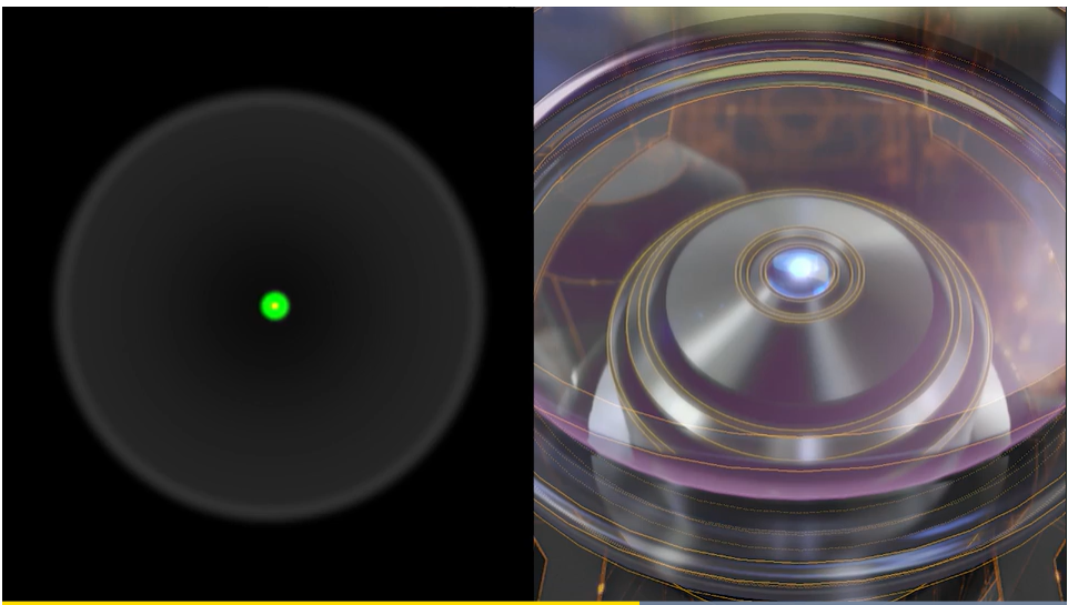
내장된 센서 모음이 현미경의 다양한 부품의 상태 정보를 감지하고 중계합니다. 컴퓨터로 이미지를 획득할 때 모든 상태 정보가 메타데이터에 기록되므로 획득 조건을 쉽게 불러오거나 구성상의 오류를 확인할 수 있습니다.
또한 내장된 카메라를 통해 후면 조리개를 볼 수 있어 DIC에서 위상 링 정렬 및 소광 교차를 쉽게 확인할 수 있습니다. TIRF와 같은 애플리케이션을 위해 레이저를 안전하게 정렬할 수 있습니다.
태블릿에서 현미경 상태를 볼 수 있고 현미경 전면의 상태 표시등을 기준으로 판단할 수도 있어 암실에서도 상태 판단이 가능합니다.
상태 표시등
운영 절차 지침
Ti2-E/A
Ti2의 어시스트 가이드 기능은 현미경 작동을 위한 대화형 단계별 지침을 제공합니다. 어시스트 가이드는 태블릿이나 PC에서 확인할 수 있으며 내장 센서와 내부 카메라의 실시간 데이터를 통합해 나타냅니다. 어시스트 가이드는 실험 설정 및 문제 해결을 위한 절차로 사용자를 돕기 위해 설계되었습니다.
① 필드 다이어프램 이미지를 시야의 중심으로 이동
② 광학 경로에서 베르트랑 렌즈를 제거합니다.
③ 관찰 포트 선택
오류 자동 감지
Ti2-E/A
확인 모드를 사용하면 선택된 관찰 방법에 필요한 현미경의 모든 구성 요소가 제자리에 있는지 태블릿이나 PC를 통해 쉽게 확인할 수 있습니다.
이 기능은 필요한 관찰 방법이 달성되지 못했을 때 문제를 찾아 해결하는 시간을 줄여줍니다.
현미경을 여러 사용자가 사용하며 각 설정이 예기치 않게 변경될 가능성이 있는 환경에서 특히 유용합니다. 맞춤형 검사 절차 또한 미리 프로그래밍할 수 있습니다.
오정렬된 구성 요소 표시
직관적인 작동
Ti2는 궁극의 사용자 경험을 위해 본체의 모든 버튼과 스위치의 종류와 위치가 완전히 새롭게 설계되었습니다. 대부분의 이미징 실험이 수행되는 어두운 환경에서도 제어를 사용하기가 쉽습니다. Ti2는 직관적이고 손쉬운 사용자 인터페이스를 제공하므로 연구원이 현미경 제어가 아닌 데이터에 집중할 수 있습니다.
신중하게 설계된 현미경 제어 레이아웃
Ti2-E, Ti2-A
모든 버튼과 스위치는 제어 대상 조명 유형을 기반으로 배치됩니다. 투과형 관찰을 제어하는 버튼은 현미경의 왼쪽에 위치하고 에피 형광 관찰을 제어하는 버튼은 오른쪽에 있습니다. 일반적인 작업을 제어하는 버튼은 전면 패널에 있습니다. 이렇게 영역을 나누어 배치함으로써 암실에서 현미경을 작동할 때 필요한 기능의 위치를 쉽게 찾아 사용할 수 있습니다.

① 셔틀 스위치(Ti2-E)
형광 필터 터렛과 대물렌즈 노즈피스 같은 장치를 제어하는 셔틀 스위치가 설계에 통합되었습니다. 이 스위치는 장치를 수동으로 회전시키는 느낌을 재현해 제어에 직관성을 부여합니다 . 하나의 셔틀 스위치에 관련된 여러 장치의 작동이 통합됩니다. 예를 들어 형광 필터 터렛의 셔틀 스위치는 터렛을 회전시키고 형광 셔터를 열고 닫는 데 사용할 수 있습니다. 배리어 필터 휠과 외부 위상차 장치를 작동시키도록 이 스위치를 프로그래밍하는 것도 가능합니다.
② 프로그래밍 가능한 기능 버튼(Ti2-E/A)
편리하게 위치한 기능 버튼을 사용해 인터페이스를 사용자 정의할 수 있습니다.
셔터 및 I/O 포트를 통해 외부 장치로 신호 출력과 같은 전동 장치 제어를 포함하여 100개 이상의 기능 중에서 선택해 이미지를 획득할 수 있습니다.
각 전동 장치의 설정을 저장하여 관찰 방법을 즉시 변경할 수 있는 모드 기능도 이 버튼에 할당할 수 있습니다.
③ 초점 조절 노브(Ti2-E)
초점 가속 버튼과 PFS 결합 버튼은 초점 조절 손잡이 옆에 위치합니다. 두 버튼의 모양이 다르기 때문에 촉감으로 쉽게 식별할 수 있습니다. 포커싱 속도는 사용 중인 대물렌즈에 맞게 자동으로 조정되므로 이상적인 포커싱 속도를 유지하여 스트레스 없는 작동이 가능합니다.
조이스틱과 태블릿을 통한 직관적인 제어
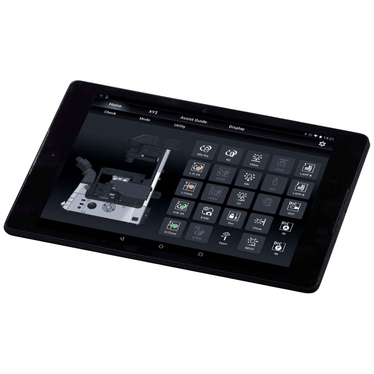
Ti2 조이스틱은 스테이지 이동을 제어할 뿐만 아니라 PFS 활동을 포함하여 현미경의 대부분의 전동 기능을 제어합니다. XYZ 좌표와 현미경 구성 요소의 상태를 표시할 수 있어 사용자가 현미경을 원격으로 제어할 수 있는 효과적인 수단을 제공합니다. 무선 LAN으로 현미경에 연결된 태블릿에서 Ti2의 전동 기능을 제어할 수 있으며 현미경 제어를 위한 다양한 그래픽 인터페이스가 제공됩니다.
Ti2 Control: 스마트폰 및 태블릿 전용 애플리케이션
Ti2-E의 설정 및 제어와 Ti2-A의 설정, 상태 표시 및 작동 가이드를 지원합니다.
다운로드
Ti2 Control 은 App Store®와 Google Play™에서 무료로 다운로드할 수 있습니다.
iPhone®, iPad® or Android™ 같은 기기에 다운로드하십시오. 호환 기기에 대한 내용은 아래의 "작동 조건" 섹션을 참조하십시오.
작동 조건
iOS 기기: iOS 10.3 이상이 설치된 기기(iPhone 7 이상, iPad Air 2 이상)
Android™ devices: devices equipped with Android 5.1 or later
Apple®, App Store®, Apple 로고,, iPhone® 및 iPad®는 미국을 비롯한 여러 국가에서 Apple Inc.의 등록 상표입니다.
iOS 상표는 미국을 비롯한 여러 국가에서 Cisco의 허가를 받아 사용됩니다.
iPhone 상표는 Aiphone Co. Ltd.의 허가를 받아 사용됩니다.
Android™ 와 Google Play™는 Google Inc.의 등록 상표입니다.
스펙
