-
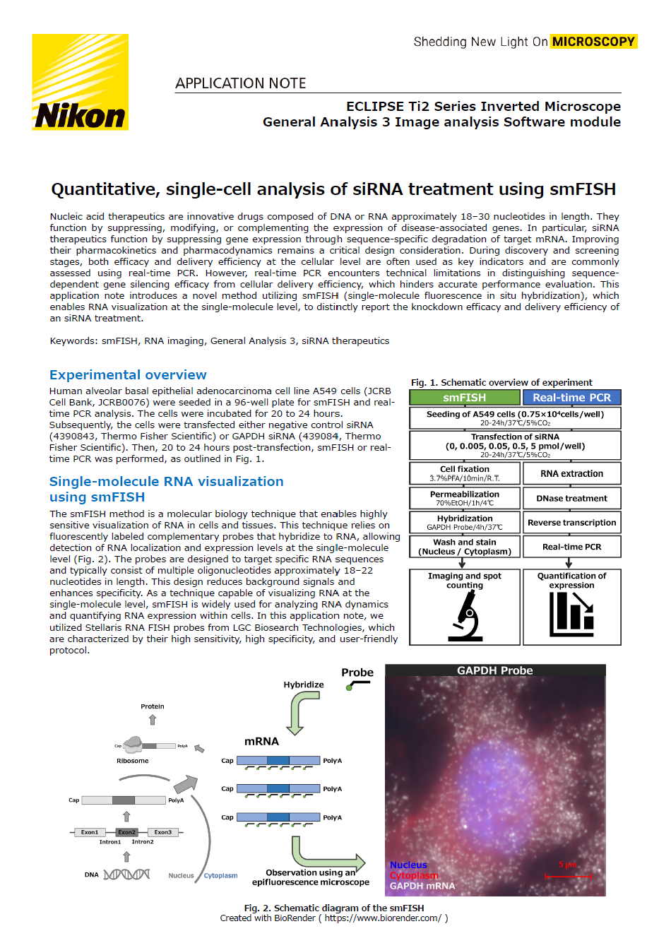
Quantitative, single-cell analysis of siRNA treatment using smFISH
-
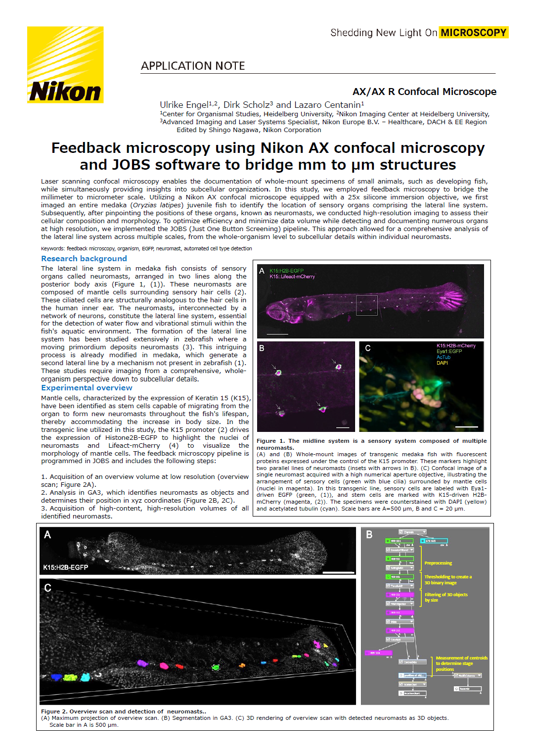
Feedback microscopy using Nikon AX confocal microscopy and JOBS software to bridge mm to µm structur
-
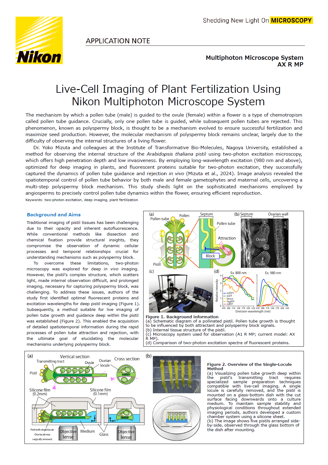
Live-Cell Imaging of Plant Fertilization Using Nikon Multiphoton Microscope System
-
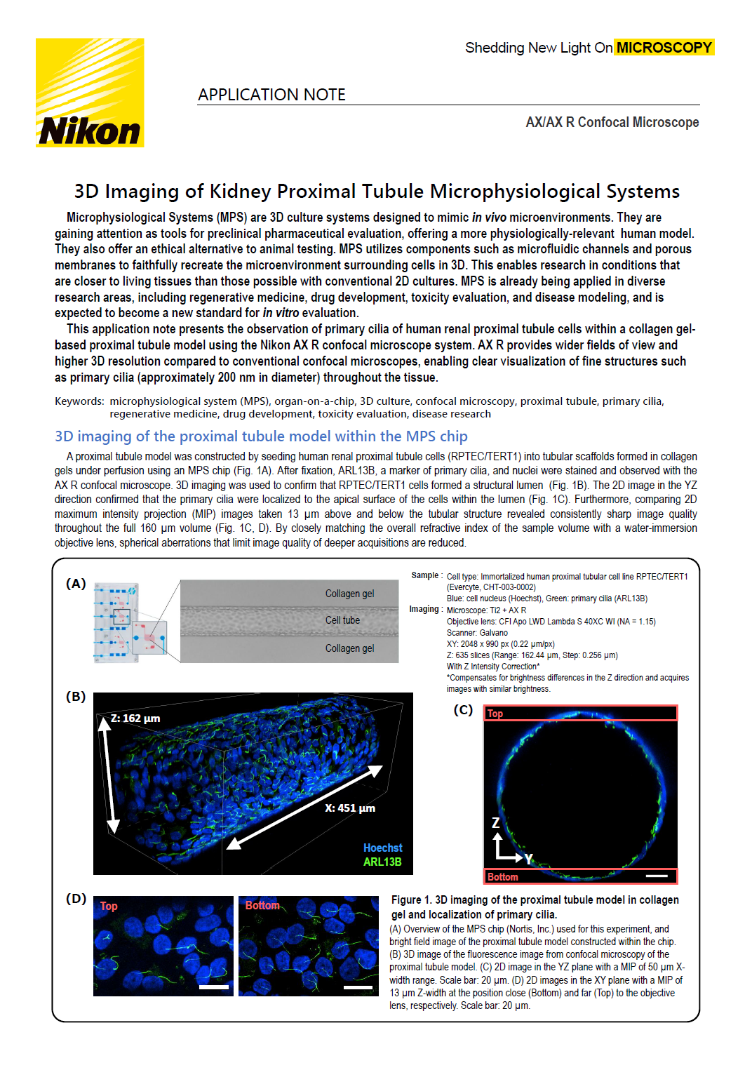
3D Imaging of Kidney Proximal Tubule Microphysiological Systems
-
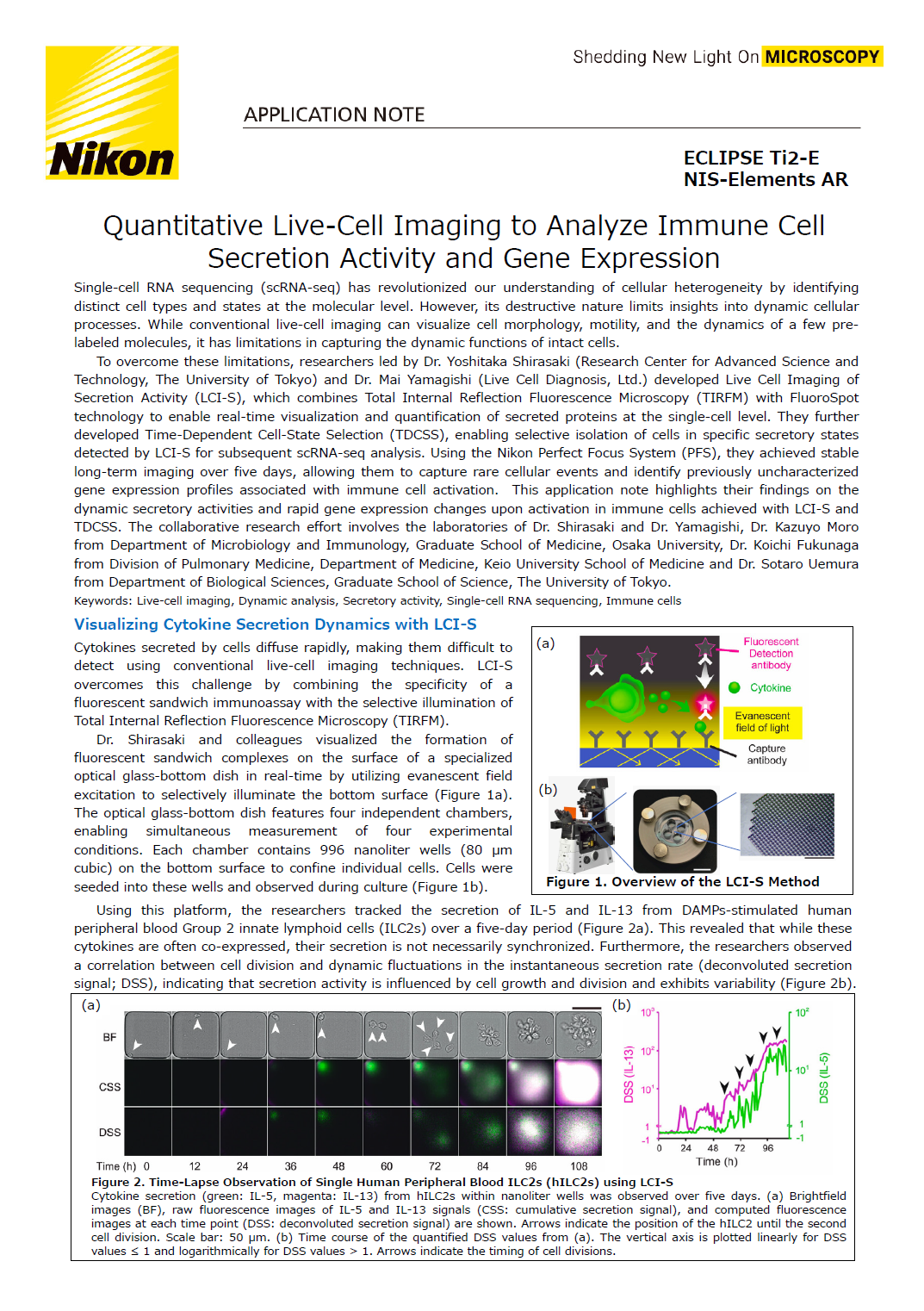
Quantitative Live-Cell Imaging to Analyze Immune Cell Secretion Activity and Gene Expression
-
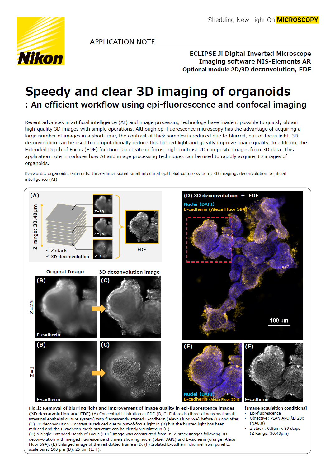
Speedy and clear 3D imaging of organoids
-
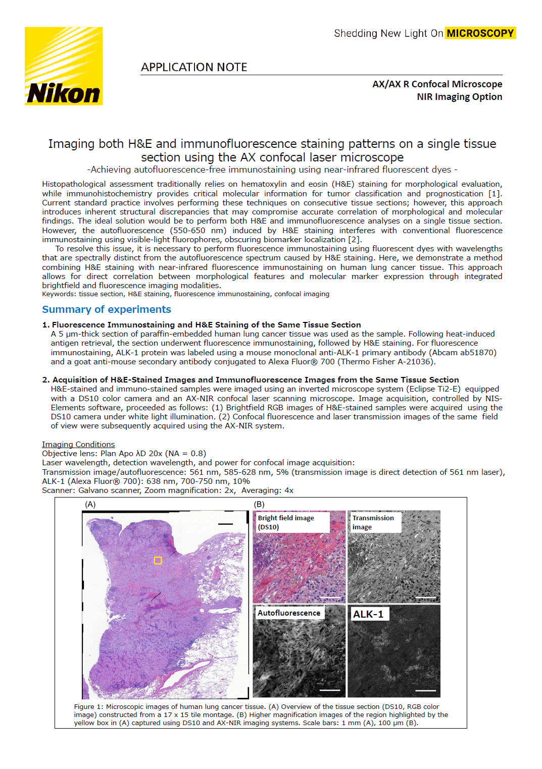
Imaging both H&E and immunofluorescence staining patterns on a single tissue section using the AX
-
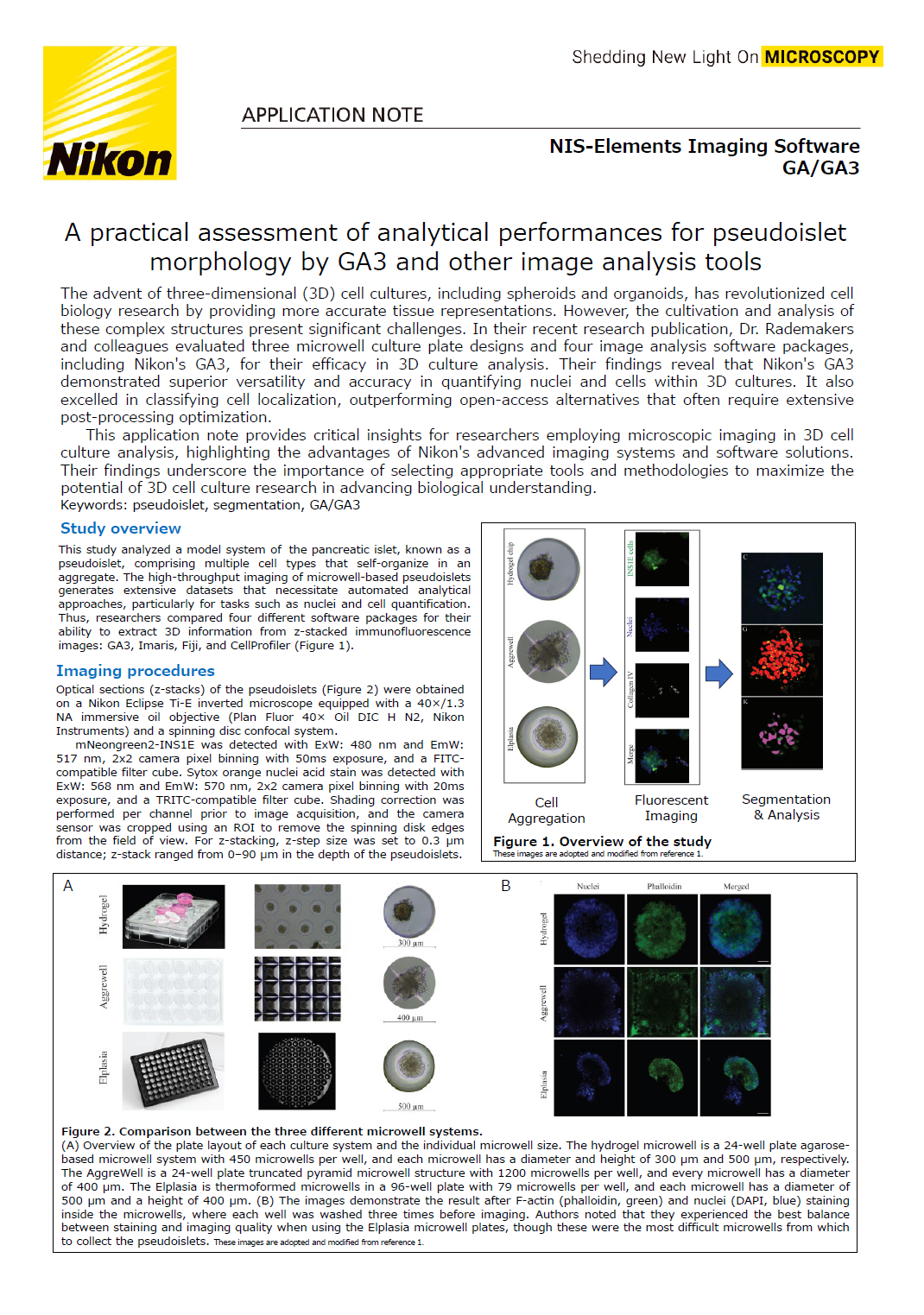
A practical assessment of analytical performances for pseudoislet morphology by GA3 and other image
-
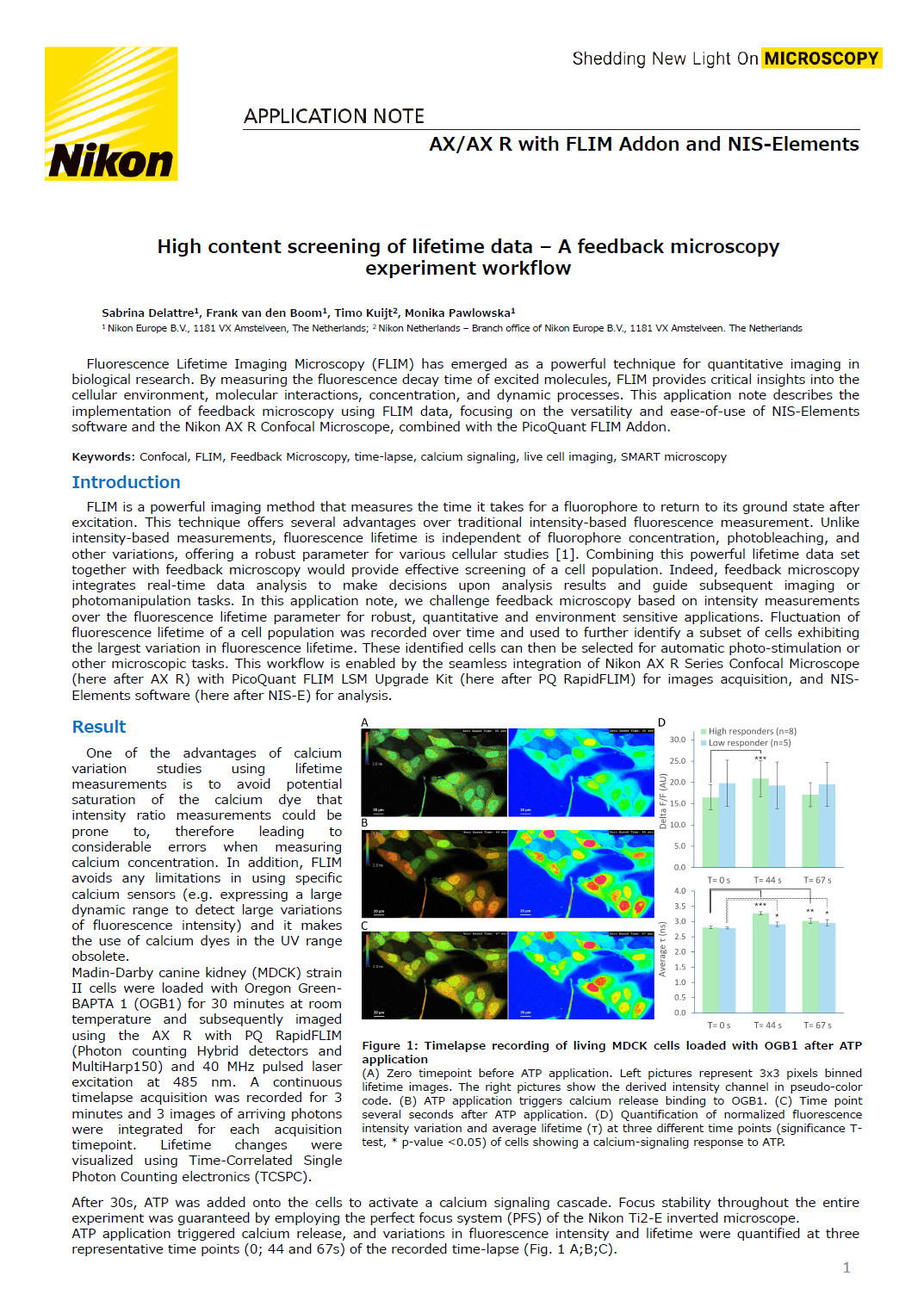
High content screening of lifetime data – A feedback microscopy experiment workflow
-
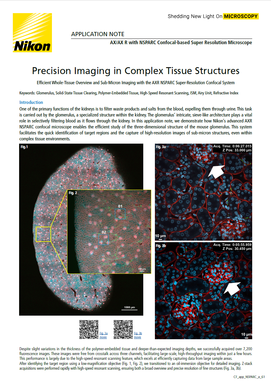
Precision Imaging in Complex Tissue Structures
-
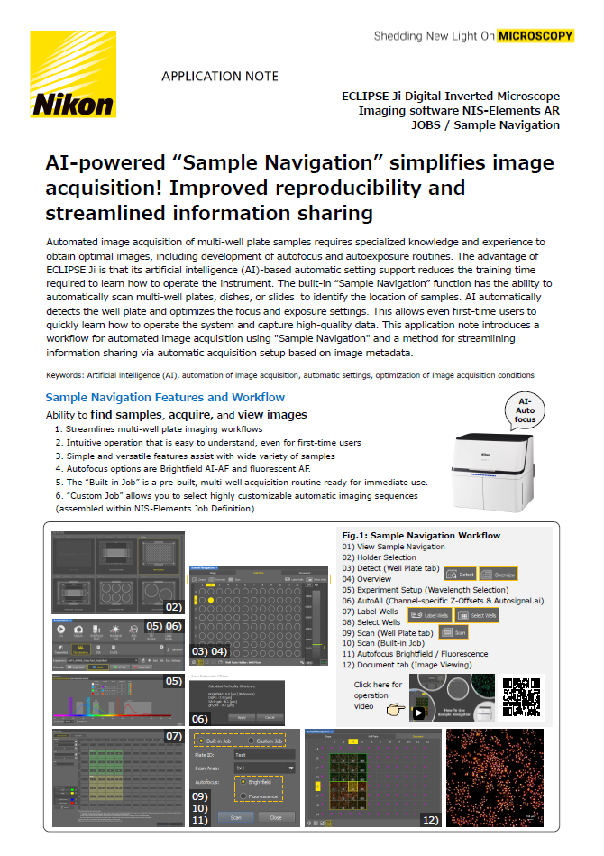
AI-powered “Sample Navigation” simplifies image acquisition! Improved reproducibility and streamline
-
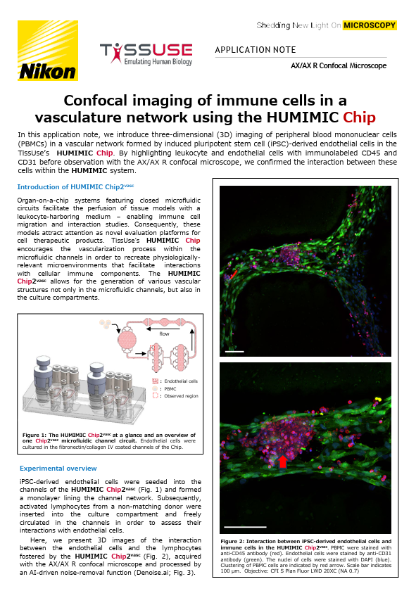
Confocal imaging of immune cells in a vasculature network using the HUMIMIC Chip
-
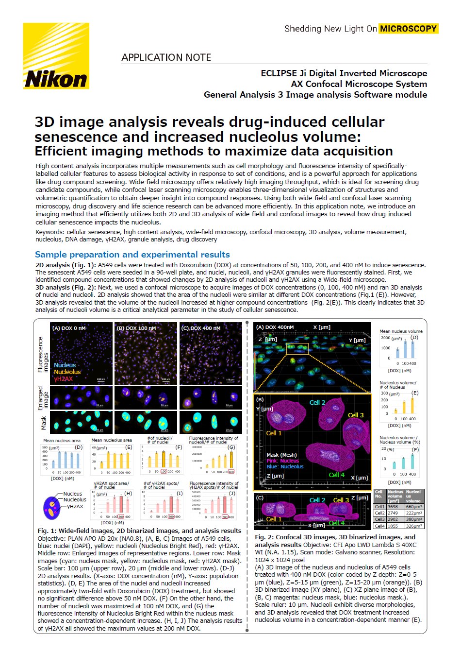
3D image analysis reveals drug-induced cellular senescence and increased nucleolus volume: Efficient
-
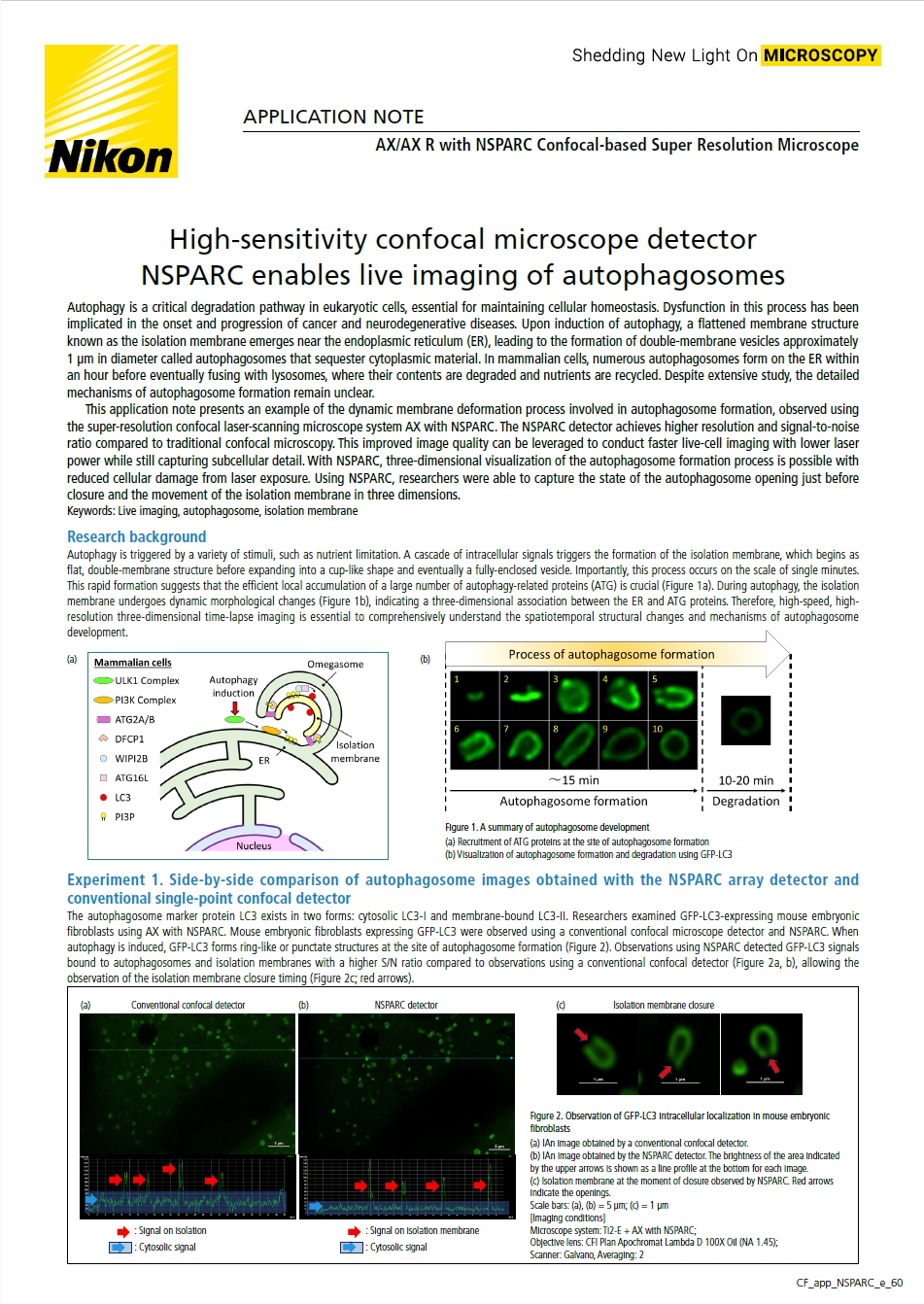
High-sensitivity confocal microscope detector NSPARC enables live imaging of autophagosomes