Speedy and clear 3D imaging of organoids.
An efficient workflow using epi epi-fluorescence and confocal imaging
Recent advances in artificial intelligence (AI) and image processing technology have made it possible to quickly obtain high -quality 3D images with simple operations. Although epi epi-fluorescence microscopy has the advantage of acquiring a large number of images in a short time, the contrast of thick samples is reduced due to blurred, out out-of -focus light. 3D deconvolution can be used to computationally reduce this blurred light and greatly improve image quality. In addition, the Extended Depth of Focus (EDF) function can create in in-focus, high high-contrast 2D composite images from 3D data. This application note introduces how AI and image processing techniques can be used to rapidly acquire 3D images of organoids.
Keywords: organoids, enteroids, three three-dimensional small intestinal epithelial culture system, 3D imaging, deconvolution, artificial intelligence (AI)
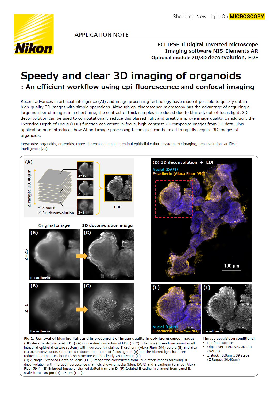
Fig.1: Removal of blurring light and improvement of image quality in epi-fluorescence images (3D deconvolution and EDF)
(A) Conceptual illustration of EDF. (B, C) Enteroids (three-dimensional small intestinal epithelial culture system) with fluorescently stained E-cadherin (Alexa Fluor 594) before (B) and after (C) 3D deconvolution. Contrast is reduced due to out-of-focus light in (B) but the blurred light has been reduced and the E-cadherin mesh structure can be clearly visualized in (C).
(D) A single Extended Depth of Focus (EDF) image was constructed from 39 Z-stack images following 3D deconvolution with merged fluorescence channels showing nuclei (blue: DAPI) and E-cadherin (orange: Alexa Fluor 594). (E) Enlarged image of the red dotted frame in D, (F) Isolated E-cadherin channel from panel E.
Scale bars: 100 μm (D), 25 μm (E, F).
[Image acquisition conditions]
• Epi-fluorescence
• Objective: PLAN APO 20x (NA 0.8)
• Z stack : 0.8um x 39 steps (Z Range:30.40um)
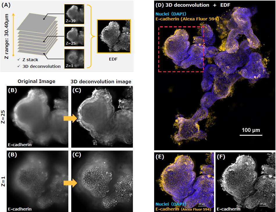
An efficient workflow combining epi-fluorescence and confocal imaging
Autosignal.ai uses AI to determine the optimal exposure conditions for observing your sample, allowing you to set the imaging conditions with a single click. Epi-fluorescence microscopy allows for quick identification of regions of interest, while switching to confocal to acquire 3D images of the regions of interest provides a sharper picture and deeper understanding of the three-dimensional structure.
Experiment 1: Epi-fluorescence imaging using 3D deconvolution and EDF
39 Z-stack images were acquired in just 18 seconds using two fluorescence wavelengths. In this experiment, it took approximately 3 minutes and 30 seconds from acquiring an image of the entire slide using "sample navigation", precisely centering the sample, acquiring images across the ~30 μm thick organoids, and the completion of image processing. This quick workflow can improve the throughput of acquiring clear images of multiple organoid samples.
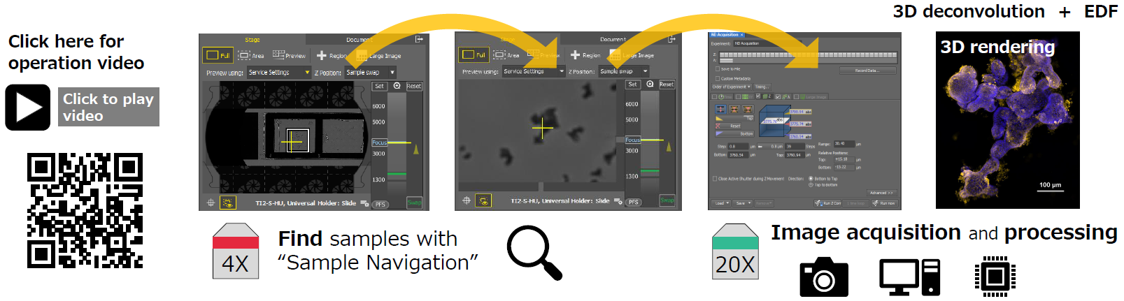
■ Imaging flow using "Sample Navigation"
1. "Sample navigation" captures the entire slide area to help locate the sample.
2. Use the 4x objective to find the organoid of interest, then switch to the 20x objective and acquire Z-stack images with the "ND Acquisition" function.
3. Process the Z-stack images using 3D deconvolution to reduce blurred light, and then construct a single all-in-focus image using EDF
■ Image acquisition and processing time: 1 minute 38 seconds in total
• Image acquisition time: 18 seconds (Magnification: 20X, Z-stack: 0.8μm x 39 steps (Z Range:30.40μm)
• Image processing time: 1 minute 20 seconds (3D deconvolution by Richardson Lucy: 52 seconds, EDF: 28 seconds). *Using NVIDIA Quadro RTX 4500
Experiment 2: Epi-fluorescence imaging using a high NA objective and 2D deconvolution
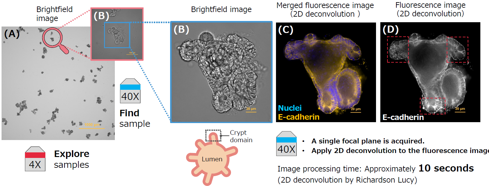
Fig. 2: Epi-fluorescence imaging and 2D deconvolution of enteroids with crypt domains.
(A) Brightfield image of enteroids, objective: PLAN APO AD 4x (NA 0.2). (B) Brightfield image of enteroids with crypt domains, Objective: PLAN APO AD 40x (NA 0.95), (C) Merged fluorescence image processed with 2D deconvolution (Nuclei (blue: DAPI), E-cadherin (orange: Alexa Fluor 594)). (D) Isolated E-cadherin channel from panel C. The high-NA PLAN APO AD 40x (NA0.95) objective produces images with a shallow depth of focus. In effect, a thinner section of the sample volume is observable. Processing this image with 2D deconvolution enables clear observation of the crypt domains (the red dotted frame in D). Scale bars: 1000 μm (A), 20 μm (B, C, D).
Experiment 3: Confocal imaging using a high magnification silicon immersion objective
In deep regions of the enteroid that have not undergone tissue clearing, light is attenuated and the image becomes darker. The Denoise.ai feature uses artificial intelligence (AI) to remove shot noise from resonant scanning confocal images based on a trained neural network to improve image quality. By applying Denoise.ai to images acquired with a high-speed resonant scanner, shot noise was removed, and image quality was greatly improved even in dark, deep regions (Fig. 3 D-1, D-2). This allowed us to clearly observe the region around the Paneth cells in the crypt domain.
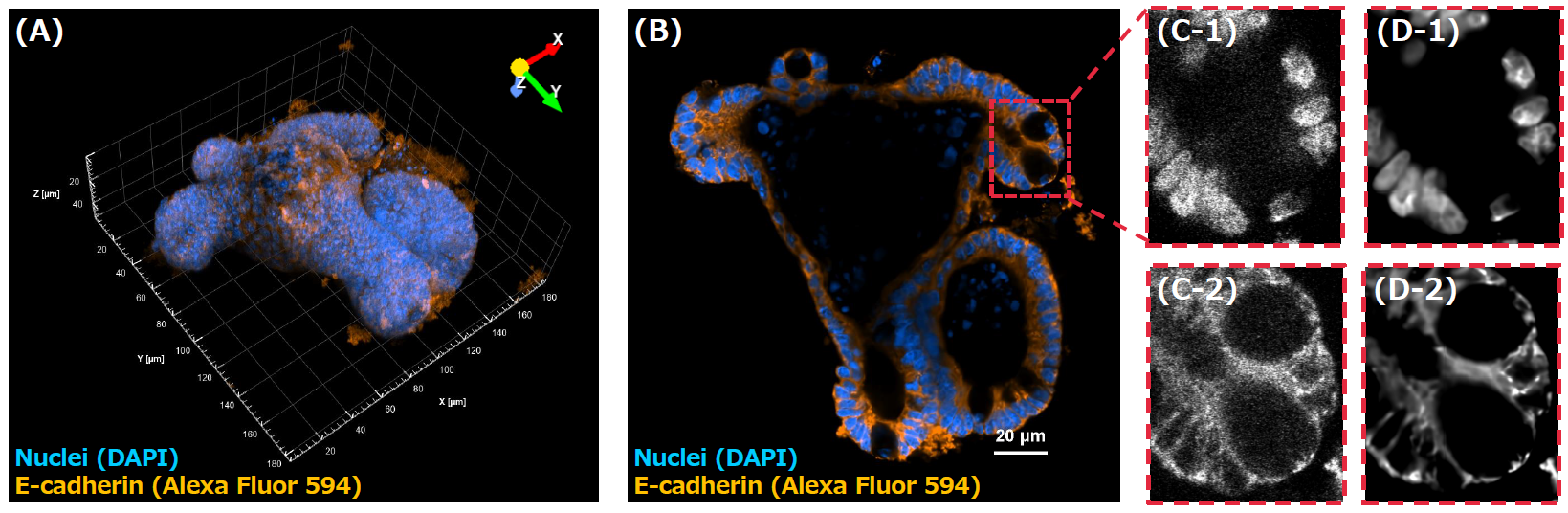
Fig.3: Confocal imaging of enteroids with crypt domains with improved image quality from Denoise.ai
(A, B) Confocal images of nuclei (blue: DAPI) and E-cadherin (orange: Alexa Fluor 594) fluorescently stained enteroids. Scale bar: 20 μm (B)
(A) Three-dimensional volumetric reconstruction of enteroids composed of 227 images (Z range: 53.30um).
(B) Optically sectioned image at Z=123 (Z depth: 28.77 μm), (C, D) Enlarged image of the red dotted frame region in B.
(C-1, C-2) Original images before applying Denoise.ai, (D-1, D-2) Images after applying Denoise.ai,
(D-1) Confocal images of nuclei (DAPI)
(D-2) Confocal images of E-cadherin (Alexa Fluor 594)
[Image acquisition conditions]
• Objective: Plan Apo Lambda S 60XC Sil (NA1.3)
• Zoom: 1.3x
• Resonant scanner
• 2K x 2K pixels
• Averaging: 4x
• Z range : 53.30um (Z Step 0.236um x 227 steps)
Summary
● Advantages of AI
✓ Easy to use: Automates image acquisition and processing tasks to make capturing clear images easier.
● Advantages of epi-fluorescence microscopy combined with image processing techniques
✓ Speedy image acquisition: Images are acquired using a camera at high frame rates, allowing you to quickly identify areas of interest.
✓ Improved image quality by removing blurred light: 2D/3D deconvolution removes out-of-focus blurring light to provide images with sharper details.
● Advantages of confocal microscopy and image processing
✓ Acquire 3D images: Optically-sectioned views within volumetric images offer a deeper understanding of three-dimensional structures.
✓ Thick samples: Confocal microscopy blocks out-of-focus light and provides sharp images with high resolution.
✓ Remove noise: Denoise.ai removes shot noise to improve rapid or dim-sample imaging.
● Advantages of combining epi-fluorescence and confocal imaging
✓ Efficient region-of-interest identification: Fast image acquisition with epi-fluorescence imaging allows you to quickly identify regions of interest. Then, confocal microscopy can be used to acquire 3D images and observe finer details. Combining these modalities improves workflows by shortening the total time from identifying an area of interest to detailed observation.
Related Application Notes
1. Selecting the Right Objectives - Bright, Sharp Imaging of Structures down to Deep Areas
2. Live Imaging of Paneth Cell Secretory Responses in Innate Immunity by Using Three-Dimensional Culture of Small Intestinal Epithelial Cells
3. The Advantages of Resonant Scanning with Ultra Short Laser Exposure Times in Live Imaging
4. Nikon NIS-Elements Denoise.ai Software: utilizing deep learning to denoise confocal data
Acknowledgements
Nikon Corporation expresses its sincere thanks to Dr. Yuki Yokoi, Dr. Kiminori Nakamura in the Innate Immunity Laboratory, Faculty of Advanced Life Science, Hokkaido University, for providing us with the specimens.
Author Information
Chiharu Kobayashi
Nikon Corporation, Tokyo, Japan
Product Information
ECLIPSE Ji Digital Inverted Microscope with AX Confocal Microscope System
The ECLIPSE Ji is capable of fast, high-resolution image acquisition using its built-in camera and epi-fluorescence light source. This digital inverted microscope is optimized for 2D high content analysis, with the ability to upgrade to confocal when required. From drug screening using fast wide-field imaging to understanding three-dimensional structures using confocal images, you can obtain seamless results with this single unit.

스펙