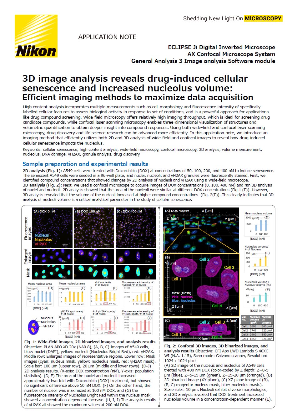
3D image analysis reveals drug-induced cellular senescence and increased nucleolus volume: Efficient imaging methods to maximize data acquisition
High content analysis incorporates multiple measurements such as cell morphology and fluorescence intensity of specifically-labelled cellular features to assess biological activity in response to set of conditions, and is a powerful approach for applications like drug compound screening. Wide-field microscopy offers relatively high imaging throughput, which is ideal for screening drug candidate compounds, while confocal laser scanning microscopy enables three-dimensional visualization of structures and volumetric quantification to obtain deeper insight into compound responses. Using both wide-field and confocal laser scanning microscopy, drug discovery and life science research can be advanced more efficiently. In this application note, we introduce an imaging method that efficiently utilizes both 2D and 3D analysis of wide-field and confocal images to reveal how drug-induced cellular senescence impacts the nucleolus.
Keywords: cellular senescence, high content analysis, wide-field microscopy, confocal microscopy, 3D analysis, volume measurement, nucleolus, DNA damage, γH2AX, granule analysis, drug discovery
스펙