High-sensitivity confocal microscope detector NSPARC enables live imaging of autophagosomes.
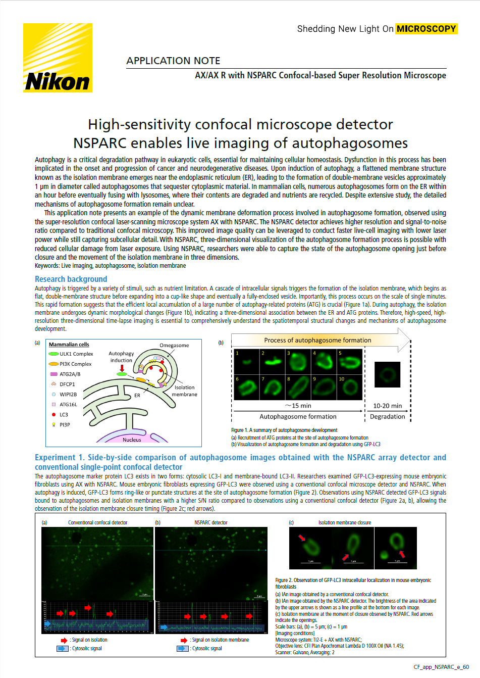
Autophagy is a critical degradation pathway in eukaryotic cells, essential for maintaining cellular homeostasis. Dysfunction in this process has been implicated in the onset and progression of cancer and neurodegenerative diseases. Upon induction of autophagy, a flattened membrane structure known as the isolation membrane emerges near the endoplasmic reticulum (ER), leading to the formation of double-membrane vesicles approximately 1 μm in diameter called autophagosomes that sequester cytoplasmic material. In mammalian cells, numerous autophagosomes form on the ER within an hour before eventually fusing with lysosomes, where their contents are degraded and nutrients are recycled. Despite extensive study, the detailed mechanisms of autophagosome formation remain unclear.
This application note presents an example of the dynamic membrane deformation process involved in autophagosome formation, observed using the super-resolution confocal laser-scanning microscope system AX with NSPARC. The NSPARC detector achieves higher resolution and signal-to-noise ratio compared to traditional confocal microscopy. This improved image quality can be leveraged to conduct faster live-cell imaging with lower laser power while still capturing subcellular detail. With NSPARC, three-dimensional visualization of the autophagosome formation process is possible with reduced cellular damage from laser exposure. Using NSPARC, researchers were able to capture the state of the autophagosome opening just before closure and the movement of the isolation membrane in three dimensions.

Keywords: Live imaging, autophagosome, isolation membrane
스펙