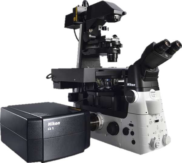
A confocal microscope that captures images with a 25 mm field of view, nearly twice the area of conventional point scanners.
Capturing images of large samples such as tissues, organs and live model organisms requires both extending the detectable area of cellular responses and increasing image capture speed. The A1 HD25/A1R HD25 confocal microscope has the largest field of view (25 mm) in its field, enabling users to expand the limits of scientific research.
See more than before in confocal resolution
When combined with the Ti2-E inverted microscope, the imaging area of the A1 HD25/A1R HD25 is nearly twice the conventional FOV of 18 mm, enabling the user to obtain significantly more data by capturing more of the specimen in each shot.
New 25mm FOV
Fewer total images required for large high-resolution image stitching
The large 25 mm FOV of the A1 HD25/A1R HD25 reduces both the required number of images for stitching large images and image acquisition times, enabling efficient, high-throughput imaging of even large-scale samples.
The number of required images can be greatly reduced, in particular for large 3D (XYZ) image stitching.
-
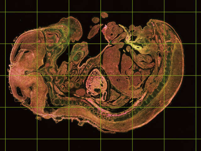
FOV 25 of A1 HD25/A1R HD25: a total of 24 frames
-
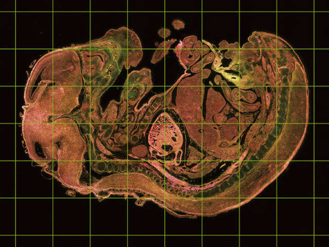
Conventional FOV 18: a total of 48 frames
Increase analytical throughput without compromising image resolution
The combination of a high-speed resonant scanner and large field of view forms an ideal platform for high-resolution screening assays. It dramatically reduces the time needed to analyze multiple samples and conditions.
-
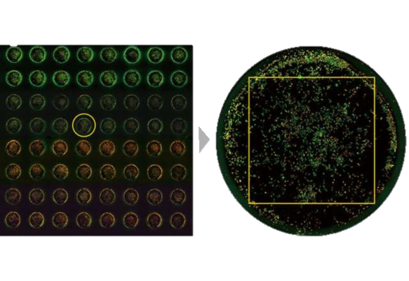
Left: A well in the 96-well plate is selected
Right: The large FOV enables acquisition of entire we -
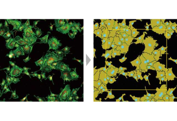
Large FOV enables measurement of larger areas and high-throughput analysis
High-speed, high-definition resonant scanner
High definition imaging up to 1K x 1K
1024 x 1024 pixels enables acquisition of high-resolution, high-quality images at lower magnifications, enabling compatibility with a wide range of samples.
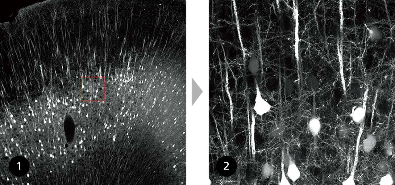
① 1 x zoom (1024 x 1024 pixels)
② 6 x zoom (1024 x 1024 pixels)
Comparison of a large FOV image and 6X zoomed image (1024 x 1024 pixels) of fine structures in a 2 mm brain slice of H-line mouse cleared with RapiClear1.52, SunJinLab.
Image courtesy of: Drs. Ryosuke Kawakami, Kohei Otomo, and Tomomi Nemoto, Research Institute for Electronic Science, Hokkaido University
Low phototoxicity for live cells
High speed imaging capability up to 720 fps, in combination with a large field of view, dramatically increases imaging throughput.
This scanning method reduces the exposure time of the sample to excitation light, minimizing phototoxicity and photobleaching.
Galvano Scanner
Resonant Scanner
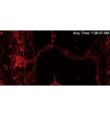
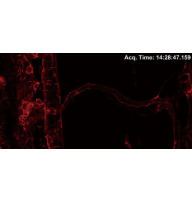
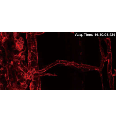
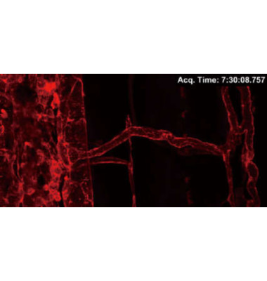
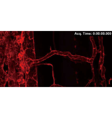
Comparison of photobleaching of fluorescent proteins when images are acquired using both galvano and resonant scanners.
3D time-lapse images of trunk vasculature in zebrafish larva expressing LIFEACT-mCherry (probe for F-actin) in endothelial cells were acquired every 30 minutes over a period of 15 hours using a galvano scanner (average of 2 images) and a resonant scanner (average of 64 images).
1024 x 512 pixels, 2X zoom, 100 Z-stack images
Note that photobleaching of LIFEACT-mCherry was dramatically suppressed using the resonant scanner.
Image courtesy of: Shinya Yuge Ph.D., and Shigetomo Fukuhara, Ph.D., Department of Molecular Pathophysiology, Institute of Advanced Medical Sciences, Nippon Medical School
스펙
