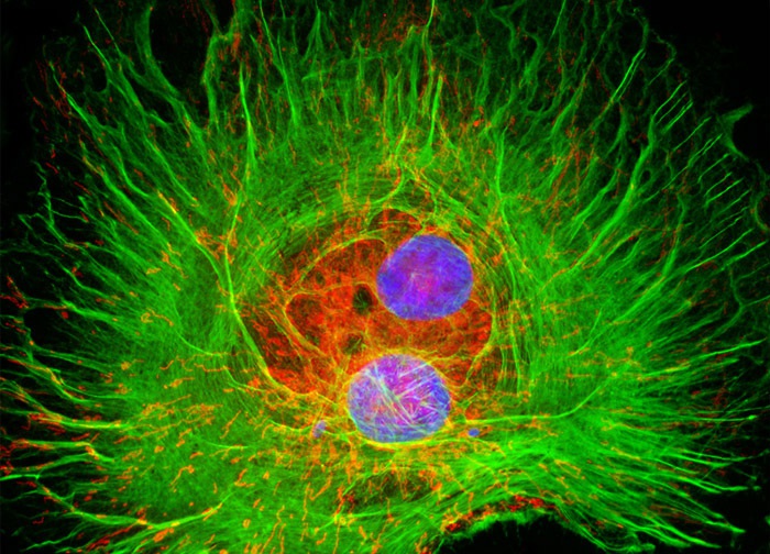
Normal African Green Monkey Kidney Fibroblast Cells
The CV-1 cell line is a widely utilized fibroblast line that was established in the mid-1960s.
When the line was first initiated, CV-1 cells were primarily used in investigations of Rous sarcoma virus (RSV).
More recently, the CV-1 line has garnered a significant amount of usage as a host for acquired immunodeficiency disease (AIDS) research.
The cells are also often employed in transfection experiments with simian virus 40 (SV40) and recombinant plasmid vectors.
CV-1 cells demonstrate susceptibility to a variety of viruses, including herpes simplex, Eastern and Western equine encephalitis, poliovirus 1, California encephalitis, and simian virus 40.
The intracellular relationship between the cytoskeletal filamentous actin network and mitochondria present in a culture of CV-1 fibroblast cells (illustrated above) was visualized with the use of the probes Alexa Fluor 488 conjugated to phalloidin (yielding green fluorescence emission) and MitoTracker Red CMXRos. Cell nuclei were counterstained with DAPI (blue emission).
Images were recorded in grayscale with a 12-bit digital camera coupled to either a Nikon E-600 or Eclipse 80i microscope equipped with bandpass emission fluorescence filter optical blocks.
During the processing stage, individual image channels were pseudocolored with RGB values corresponding to each of the fluorophore emission spectral profiles.

Normal African Green Monkey Kidney Fibroblast Cells (CV-1 Line)
The culture of African green monkey kidney cells presented in the digital image above was immunofluorescently labeled with primary anti-tubulin mouse monoclonal antibodies followed by goat anti-mouse secondary antibody fragments (Fab) conjugated to Rhodamine Red-X.
In addition, the specimen was subsequently counterstained for DNA in the cell nucleus with DAPI. Images were recorded in grayscale with a 12-bit digital camera coupled to either a Nikon E-600 or Eclipse 80i microscope equipped with bandpass emission fluorescence filter optical blocks. During the processing stage, individual image channels were pseudocolored with RGB values corresponding to each of the fluorophore emission spectral profiles.
Normal African Green Monkey Kidney Fibroblast Cells (CV-1 Line)
he culture of African green monkey kidney (CV-1) cells that is presented in the digital image above was labeled with SYTOX Green and Alexa Fluor 350 conjugated to phalloidin, which target nuclear DNA and filamentous actin, respectively.
The specimen was also stained for mitochondria with MitoTracker Red CMXRos. Images were recorded in grayscale with a 12-bit digital camera coupled to either a Nikon E-600 or Eclipse 80i microscope equipped with bandpass emission fluorescence filter optical blocks.
During the processing stage, individual image channels were pseudocolored with RGB values corresponding to each of the fluorophore emission spectral profiles.
Normal African Green Monkey Kidney Fibroblast Cells (CV-1 Line)
The microtubules present in the log phase culture of CV-1 cells featured in the digital image presented above were immunofluorescently labeled with primary anti-tubulin mouse monoclonal antibodies followed by goat anti-mouse Fab fragments conjugated to Rhodamine Red-X. In addition, the culture was stained with Hoechst 33258, which selectively binds to DNA in cell nuclei. Images were recorded in grayscale with a 12-bit digital camera coupled to either a Nikon E-600 or Eclipse 80i microscope equipped with bandpass emission fluorescence filter optical blocks. During the processing stage, individual image channels were pseudocolored with RGB values corresponding to each of the fluorophore emission spectral profiles.
Normal African Green Monkey Kidney Fibroblast Cells (CV-1 Line)
The mitochondria present in the culture of monkey kidney cells (CV-1 line) featured in the digital image above were fluorescently labeled with MitoTracker Red CMXRos, a derivative of X-rosamine.
In addition, the culture was labeled for the cytoskeletal F-actin network and DNA in the cell nucleus with Alexa Fluor 633 conjugated to phalloidin and SYTOX Green, respectively.
Images were recorded in grayscale with a 12-bit digital camera coupled to either a Nikon E-600 or Eclipse 80i microscope equipped with bandpass emission fluorescence filter optical blocks.
During the processing stage, individual image channels were pseudocolored with RGB values corresponding to each of the fluorophore emission spectral profiles with the exception of Alexa Fluor 633, which was pseudocolored blue.
Normal African Green Monkey Kidney Fibroblast Cells (CV-1 Line)
In order to visualize lectin binding to the Golgi complex in CV-1 cells, the adherent culture illustrated above was treated with wheat germ agglutinin conjugated to Oregon Green 488.
The cells were subsequently counterstained with Alexa Fluor 568 conjugated to phalloidin to localize the filamentous actin network, and the nucleic acid stain DAPI to label DNA in the nucleus.
Images were recorded in grayscale with a 12-bit digital camera coupled to either a Nikon E-600 or Eclipse 80i microscope equipped with bandpass emission fluorescence filter optical blocks.
During the processing stage, individual image channels were pseudocolored with RGB values corresponding to each of the fluorophore emission spectral profiles.
스펙
